Nobel implants and MRI — what to tell the radiographer
Your implants are MR Conditional, not MR Safe — and one configuration has not been evaluated at all
Short version
You can have an MRI scan with dental implants. They are labelled MR Conditional, which means safe under stated conditions rather than safe in all circumstances.
Tell the radiology department you have dental implants before the scan, and tell them which system. There is one specific configuration — more than two zygomatic implants — where the honest answer is that nobody has evaluated it, and that is worth knowing in advance rather than at the door.
MR Safe, MR Conditional, MR Unsafe
Three labels, and the middle one is the one that applies here.
MR Safe — poses no known hazard in any MR environment. Typically non-metallic items.
MR Conditional — safe within specified conditions: field strength, scan duration, energy deposition limits. Outside those conditions, safety is not established.
MR Unsafe — must not enter the scanner room.
Dental implants sit in the middle category. That is normal for implanted metal and it is not cause for alarm; it simply means the conditions matter.
The published conditions
Nobel Biocare’s labelling gives the following:
- Scanning at 1.5 tesla or 3 tesla
- Within a stated spatial field gradient limit
- Within stated SAR (specific absorption rate) limits — the measure of energy deposited in tissue
- A continuous scan period of 15 minutes
- Image artefact extending up to approximately 3 cm from the implant
Most routine clinical MRI runs within these parameters. Your radiographer knows the machine’s field strength and SAR settings; what they need from you is the fact that implants are present and, ideally, which ones.
The artefact matters as much as the safety
A three-centimetre artefact is substantial, and this is the part that affects diagnosis rather than risk.
Metal distorts the local magnetic field, and the resulting void obscures anatomy near the implant. For a brain or spine scan this is usually irrelevant. For imaging of the jaw, face, sinuses, salivary glands or the floor of the mouth it can obscure the exact region being examined.
Tell the radiologist before the scan is planned, not afterwards. There are sequences that reduce metal artefact, and knowing implants are present lets them choose one. It may also change whether MRI is the right imaging at all — CT or CBCT is sometimes the better answer for that region.
The zygomatic exception — read this if it applies to you
This is the single most important line in the article.
The MRI safety and compatibility of configurations containing more than two zygomatic implants has not been evaluated. Heating, migration and image artefact in such configurations are not known.
Not “is safe”. Not “is unsafe”. Not evaluated.
This matters because a quad-zygoma rehabilitation places four. If you have had full-arch treatment in a severely resorbed upper jaw, you may well be in this configuration, and it may not have been mentioned to you.
What to do:
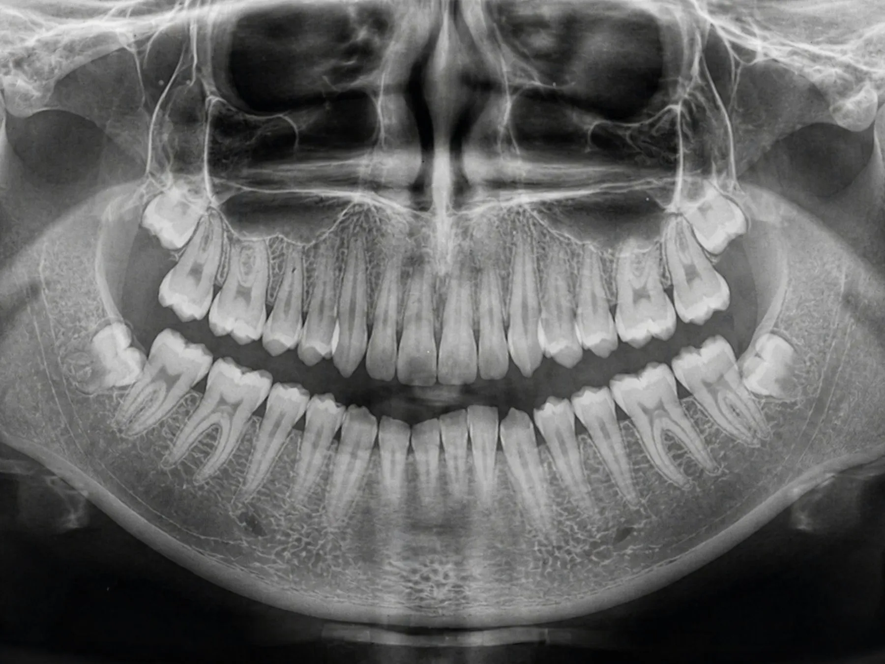
- Find out how many zygomatic implants you have. Your treatment record states it; a panoramic radiograph shows it immediately.
- Tell the radiology department the number, not just “I have implants”.
- Let the radiologist decide. They weigh the clinical need for the scan against an unquantified risk. That is their judgement to make, and they can only make it with the information.
This is not a reason to decline zygomatic treatment. It is a reason to know what you have and to carry the record.
What to take to the appointment
- Which implant system you have — manufacturer and system name
- How many implants, and how many are zygomatic
- Where they are in the mouth
- What the restoration is made of — titanium framework, zirconia, cobalt-chromium
- Your implant passport or treatment record if you have one
If you do not have that information, a panoramic radiograph taken by any dentist gives the radiology department most of it, and there are ways to identify an unknown implant from an image.
Removable and fixed work
Two practical notes:
Removable prostheses come out — dentures, overdentures, anything you take out yourself should be removed before the scan, and the department will normally ask.
Fixed work stays in. A screw-retained bridge on implants is not coming out for a scan, and it does not need to. It is part of the configuration you declare.
The reasonable conclusion
Implants do not stop you having an MRI. They add a declaration you should make and, in one specific configuration, an unknown the radiologist should be allowed to weigh.
The recurring theme of this site applies here as squarely as anywhere: the record of what was placed in your jaw is worth keeping. It is the difference between “I have some implants” and a radiographer being able to plan the scan properly.